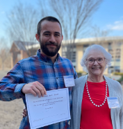
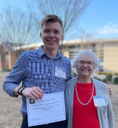
Seventy-five scientists from seven universities and the NIEHS attended the 30th Annual Meeting of the Triangle Consortium for Reproductive Biology on March 5th in Greenville, North Carolina. This in person meeting was the first in two years and allowed for welcome interpersonal interactions and sharing of outstanding science. The Campion Fund gave two awards in the form on honoraria to two junior investigators who presented their work at the meeting.

Ciro Amato, a postdoctoral fellow in Humphrey Yao’s Lab at NIEHS, was awarded the Campion Fund Award for his oral presentation entitled “A novel cell population from hindlimbs interacts with mesenchymal cells in the external genitalia to facilitate proper penis formation.” He described studies that seek to understand how the male urethra closes in the penis during fetal development. These studies are important because the incomplete closure of the urethra along the penis (hypospadias) is the second most common birth defect in the United States. This defect often requires surgical correction. If not corrected, it can lead to difficult urination and intercourse, leaving lasting physical and psychological impacts. Thus, understanding penis development has significant implications on human health.
In the male embryo, the urethra first starts to close at the base of the penis. This closure continues up the penis in a zipper-like fashion and eventually forms a tube along the length of the structure. Disruption at any time during the closure event results in hypospadias. Dr. Amato’s research found a novel population of mesenchymal cells from the hindlimb (embryonic cells that will develop into the posterior legs) that are responsible for proper penile urethra closure in the mouse. After the onset of penis formation, a population of mesenchymal cells appears near the hindlimb of the fetus. Just prior to urethra closure initiation, these hindlimb-derived cells migrate centrally and are gradually positioned bilaterally to the penile urethra. Using single cell mRNA sequencing, he discovered that these hindlimb-derived cells extensively interact with a neighboring mesenchymal cell population through genes related to extracellular matrix, cell migration, and morphogens. Removal of the hindlimb-derived cells, using a cell-type specific ablation model, resulted in severe urethra closure malformations. In many cases there was a failure to initiate the closure of the urethra. Single cell analysis of control and hind-limb ablated mice revealed transcriptomic shifts in the neighboring mesenchymal cells in the penis. These results reveal hindlimbs as a source of mesenchymal cells in the penis that are required for the initiation of urethra closure. Their role in urethra closure involves extracellular matrix gene expression in the hindlimb and neighboring cell populations inside the penis. The discovery of these hindlimb-derived cells will allow further studies to understand the biology of hypospadias.

Oleksandr Kirsanov, a student in Chris Geyer’s Lab, East Carolina University, was awarded a Campion Fund Award for his poster “Retinoic acid is dispensable for initiation and progression through male meiosis in mice.” Meiosis is a specialized form of cell division essential for sexual reproduction. In mammals, the molecular and cellular changes that germ cells must undergo to transition from mitotic spermatogonia to meiotic spermatocytes remain largely undefined. Retinoic acid (RA) has been proposed as the meiosis inducing substance (MIS) required for female and male meiotic initiation. Evidence for this presumption is based on studies of fetal gonads – meiosis is initiated in fetal ovaries but suppressed in fetal testes.
Critically, the requirement for RA in meiosis has never been formally tested because germ cell differentiation and meiotic initiation in the fetal ovary are temporally inseparable. Kirsanov used the postnatal mouse testis, where differentiation and meiotic initiation are temporally distinct (8.6 days apart), and tested the hypothesis that RA is dispensable for initiation and progression through meiosis. Complementary in vivo experimental models were designed to address the hypothesis. In the first, a “RA-sufficient” model, spermatogenesis was synchronized by administration of a potent and selective RA synthesis inhibitor; as a result, testes contained only undifferentiated spermatogonia. Then, the inhibitor was discontinued, and mice were given a single dose of exogenous RA to initiate spermatogonial differentiation. Germ cells completed differentiation, entered and completed meiosis, and formed haploid gametes at predicted timepoints. In the second, “RA-deficient” model, mice were treated as above, except the inhibitor was administered continuously. The lack of testicular RA in this model was confirmed in multiple ways, including assessment of RA-responsive gene expression, in vitro serum-free cultures, and dose-response experiments. Surprisingly, germ cells in RA-deficient testes completed differentiation, underwent meiotic DNA replication, completed the landmark cytological processes of meiosis, and formed postmeiotic haploid spermatids, all on the predicted timeline of the RA-sufficient model. To define the effect of RA deficiency on gene expression during meiotic initiation, large, highly enriched populations of premeiotic spermatogonia from RA-sufficient mice, and meiotic preleptotene spermatocytes from both models were FACS-isolated and subjected to bulk RNA-seq and shotgun proteomics.
Interestingly, analyses revealed few significant differences in expression, at the mRNA or protein levels, of characteristic meiotic genes. Taken together, their data reveal that, after initiating spermatogonial differentiation, RA is not necessary for initiation, progression through, and completion of meiosis in the male germline.


