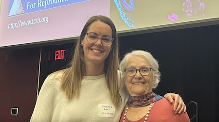
Scientists from around the country attended the Annual Meeting of the Triangle Consortium for Reproductive Biology where Phyllis Leppert was recognized in the keynote speech. We are very excited to announce our 2024 Campion Fund Awardees below!

Lenka Radonova, PhD is the recipient of the 2024 Campion Fund Award best oral presentation given at the 32nd meeting of the Triangle Consortium for Reproductive Biology held March 29, 2024. Her presentation was entitled: Mitochondria-endoplasmic reticulum domains in Ca2++ signaling at fertilization. She studied embryo development that starts at fertilization when sperm trigger oscillations in egg cytoplasmic Ca2⁺ levels. Ca2⁺ oscillations depend on Ca2⁺ release from endoplasmic reticulum (ER) stores, followed by a return to basal Ca2⁺ levels due to Ca2⁺ reuptake. Each Ca2⁺ transient stimulates mitochondrial Ca2⁺ uptake, leading to the generation of ATP that powers these pumps and other cellular activities. In somatic cells, the ER and mitochondria closely associate at mitochondria-ER-associated membrane domains (MAMs), essential for Ca2⁺ transfer from ER into mitochondria and, in turn, for cell processes like lipid synthesis, metabolism, and Ca2⁺ homeostasis. Her work demonstrated that MAMs are present in eggs. Dr. Radonova and colleagues performed focused ion beam-scanning electron microscopy with the subsequent 3-D reconstruction of the organelles of interest to determine the ER-mitochondria interactions at the ultrastructure level in mouse eggs. Their findings confirmed that the mitochondria were spherical with small immature cristae. The mitochondria were often in groups in the egg cortex, closely associated with ER tubules, suggesting close interactions of both organelles. They also observed MAMs in live mouse oocytes and eggs. There were differences in the MAM distribution in oocytes and eggs: In oocytes, the MAMs localized through the cytoplasm, with modest accumulation around the germinal vesicle. In eggs, MAMs surrounded the meiotic spindle with a “dispersal pattern” localization from the spindle through the center of the cell. In both cases, the MAM localization correlated with the mitochondria distribution in oocytes and eggs. These results provide the basis for future experiments involving the disruption of MAMs in eggs to study their impact on Ca2⁺ signaling during fertilization. Her findings will promote an understanding of the effects of health conditions and environmental factors on female reproduction.

Virginia Savy, PhD, also a member of Carmen Willliams PhD’s Laboratory at the National Institute of Environmental Health Sciences won the Campion Award for the best poster at the 32nd TCRB annual meeting. Her topic was Calcium oscillations: the egg’s elevator pitch at fertilization. Her poster presented research on the effect of calcium oscillations on the reproductive health of offspring following fertilization. In mammals, the initiation of new life involves repetitive changes in the egg’s intracellular calcium level, called calcium oscillations. These oscillations last only for a short time (about 5 h in mice) but encode essential instructions for egg activation and subsequent embryo development. She pointed out that this unique, concise, and critical discourse resembles an elevator pitch. These oscillations are susceptible to the environment and can be altered in vitro by changing the ionic composition of culture media, or in vivo by medical conditions such as obesity or inflammation. In mice, abnormal calcium oscillations at fertilization have a long-term impact on offspring growth and health. These calcium-dependent long-term outcomes raise concerns that there could be similar consequences for children conceived in vitro or from women with conditions affecting the egg’s ability to handle calcium. To study how abnormal calcium homeostasis might link to offspring health she and her colleagues studied this issue by developing a mouse model of dramatically increased calcium exposure at fertilization in vivo by knocking out (eliminating) two plasma membrane calcium pumps, called PMCA1 and PCMA 3. These PMCAs extrude excess calcium from the cytosol to maintain calcium homeostasis. PMCA1/PMCA3-null eggs (dKO) had extended elevated calcium at fertilization (10 times more than controls). When female mice without PMCA1/PMCA3 were mated with wildtype (mice with PMCA1/PCMA3) they had fewer pups per liter than control mice. Their data indicated that the pups were heavier than those of matched controls. Dr Savy and her colleagues evaluated how elevated calcium affects fertility by fertilizing dKO eggs fertilized in vitro and monitored their development. The dKO eggs were fertilized at the same rate as normal controls. However. Fewer dKO fertilized eggs reached blastocyst stage compared to controls (p≤0.007). When Dr. Savy flushed fertilized eggs from dKO females mated with WT males she found that fewer zygotes reached blastocyst stage compared to controls (p≤0.001). She also conducted experiments that demonstrated the dKO eggs with high calcium had lower global gene transcription and altered metabolism and energy production. This research suggests that altered calcium oscillations are may be detrimental in human embryo development.


