


Introduction
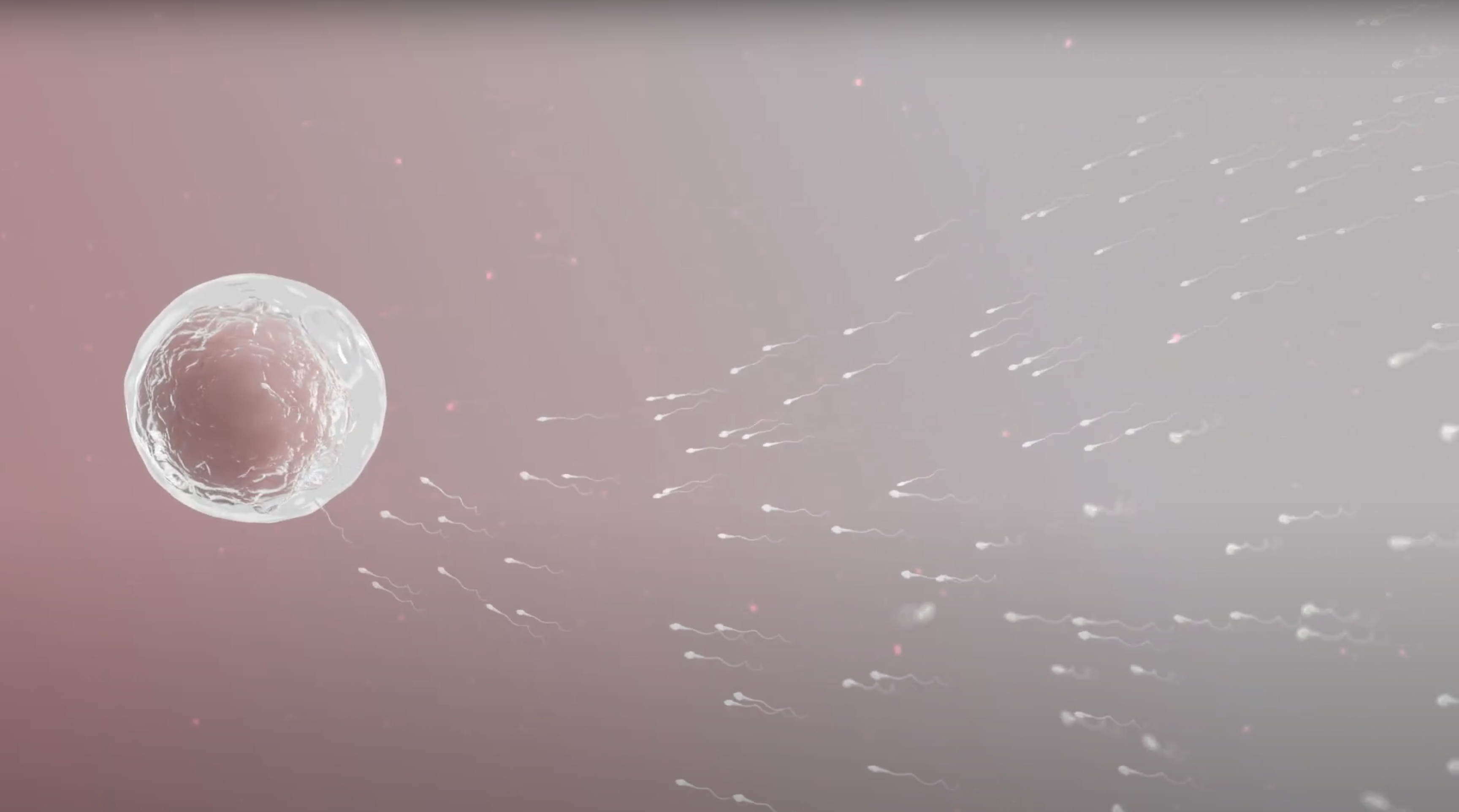
The joining together of the egg and sperm called fusion occurs over many hours. The concept of this process as being short in the minds of many persons is enhanced by the expression we use-- that the sperm penetrates the egg (oocyte). The idea of penetration is often that it occurs quickly in the way the way a spear or a bullet penetrates human tissue. Educational videos of fertilization, including ones produced by us and posted on the Campion Fund website are speeded up in terms of time giving the viewer the same impression. But this is not how fertilization works. The fusion process is complex and involves many steps in a highly orchestrated molecular mechanism involving many molecules. In humans it takes up to three days after sexual intercourse for the egg and the sperm to fuse. Fertilization is a precise biological system that takes place in an ordered time sequence. The steps of the process occur within narrow time windows. If a time window passes by, fertilization does not occur. The process of fertilization in humans is not perfect and is highly prone to errors. In fact, between 10 to 40% of all human pre-implantation embryos do not live to implantation. When this analysis of loss includes fertilization to birth the loss is 40-60%.
The fusion of the ootid and the sperm occurs in the ampulla of the Fallopian tube or oviduct near its junction with the isthmus. The ampulla is the wide part of the oviduct at its very end nearest the ovary. It is not attached to the ovary but is around it and over it like a floppy umbrella. This educational blog is meant to provide an appreciation of the complexity of human fertilization. It is not necessary to remember all the details of the process being described. However, detail is presented to provide a reference to the non-scientist who wishes to understand the process. A list for further reading is given along with a glossary. The whole stepwise process of fertilization can be confusing. However, fertilization is an awesome biological system.
Glossary
Acrosome: an organelle in the sperm head in front of the nucleus. It contains hydrolytic enzymes that allow the sperm to begin the process of fertilization.
Acrosome Reaction: is the term applied to the molecular changes that occur allowing the release of enzymes from the acrosome. The enzymes produce a hole in the zona pellucida of the secondary oocyte to allow it to pass through this thick membrane.
Capacitation: is the term applied to the changes the sperm undergoes in the female reproductive tract that enable the sperm to have the capacity to fertilize.
Cone of Attraction: The part of the secondary oocyte cytoplasm that bulges when the sperm is about to enter it. The sperm enters at that part of the secondary oocyte.
Corona Radiata: Follicular cells that surround the secondary oocyte. This is the first barrier of the cell that sperm must pass through as the processes of fertilization begin.
Cortex: The cell plasma membrane, extracellular coat and cytoskeletal structures and organelles attached to the membrane is called the cortex of the secondary oocyte.
Cortical Reaction: After one sperm enters the secondary oocyte’s cytoplasm, the perivitelline membrane forms. This membrane will keep additional sperm from entering the cytoplasm preventing polyspermy, a term applied to a situation when more than one sperm enters the secondary oocyte.
Diploid: In humans 46 chromosomes. All somatic cells are diploid.
Gamete: The reproductive cell containing a haploid number of chromosomes. In humans that number is 23.
Haploid: In humans 23 chromosomes. Haploid cells are the female and male gametes, (oocyte and sperm).
Izumo 1: is a surface protein on sperm, essential for sperm-oocyte binding.
Juno: is the receptor on the oocyte that binds Izumo 1 that occurs when the sperm enters the cytoplasm of the oocyte. After binding with Izumo 1 all other Juno receptors are lost.
Maternal Effect Genes: These genes are present in the oocyte and the fertilized cell that produce RNA and proteins prior to the initiation of the zygote gene expression.
Meiosis: The process of cell division in gametes (oocytes and sperm) that results in half of the number of chromosomes of the cells of the parents.
Mitochondria: an organelle seen in large numbers in most cells of the body where the biochemical production of energy occurs. It has two membranes; the inner membrane is folded into cristae. Mitochondria contain DNA inherited always form the mother.
Mitosis: Process of cell division in all cells except gametes that results in each new cell having a diploid number of chromosomes: in humans the number is 46.
Ootid: a haploid cell that is formed after the sperm enters the secondary oocyte. The completion of meiosis starts. The division of the ootid results in an ovum. A polar body is also created. Both are haploid cells
Ovum: The mature female gamete or final reproductive cell in the process of meiosis. It is formed when the ootid divides. This is the cell that fuses its pronucleus with the sperm pronucleus and gives rise to a zygote. It is the largest cell, along with the oocytes that precede it, in the human body and can be seen with the naked eye. It is the size of a period at the end of a sentence. This final cell forms after the sperm enters the secondary oocyte cytoplasm.
Perivitelline Space: The space between the corona radiata and the zona pellucida in the secondary oocyte.
Polar Body: A cell produced during meiosis that contains 23 chromosomes and a small amount of cytoplasm. It disintegrates soon after it is formed.
Primary Oocytes: Primary oocytes are produced in a female embryo. Primary oocytes undergo meiosis in the embryo until prophase I and then stop or arrest. They remain in this arrested state until after puberty. Thus, they live in phase I arrest for 12- 18 years depending on when puberty begins in the female child.
Pronucleus: The nuclei of the sperm and ovum are called pronuclei during fertilization just before the genetic material begin to fuse.
Secondary Oocyte: An immature ovum that is produced my meiosis shortly after ovulation. It lasts until fertilization when it progresses to an ootid and then ovum. It is a large as the ovum as it contains a large amount of cytoplasm.
Sperm: Male reproductive cell. This haploid cell in humans has a head and a tail and is highly mobile in order to traverse of swim up through the vagina, uterine cervix, uterine cavity, and part of the Fallopian Tube oviduct to meet the secondary oocyte and begin the process of fusion. Sperm have either a Y or an X chromosome and thus determine the sex of the child
Zona Pellucida: is a fibrillar network of glycoproteins bound together that surround the secondary oocyte.
Zygote: This cell is the first cell formed after complete fusion of the ovum and sperm. It contains all the genetic material necessary to form a new individual.
Zygote Activation: After the ovum and sperm pronuclei fuse and the genetic material in the nucleus are together in the new cell or zygote many changes must occur before the new cell begins transcription of DNA. The DNA does not begin to duplicate immediately. The new cell the zygote must go through a period of reprogramming to begin this process. Gradually the genes on the chromosomes are activated during a process called maternal-to-zygote transition. This enables the zygote to replace the maternal genes that initiated development.
Oocytes Mature
Oocytes begin to mature during the female menstrual cycle. Human menstrual cycles vary in length between individual women and in one woman they will vary in length throughout her reproductive life. They are considered normal if they occur most of the time every 23 to 35 days, with an average of 28 days. The cycle as most people know is under that control of female reproductive hormones. Not to get into too much detail it is very important to know or remember that primary oocytes develop in the female during the time she was an embryo-fetus in her mother’s uterus. This development involves meiosis, the process of cell division where each cell has only 23 chromosomes called haploid cells. Somatic cells are diploid cells and have 46 chromosomes. During reproduction it is important to reduce the number of chromosomes to 23 so that when the oocyte fuses with a sperm that also has 23 chromosomes, the newly formed cell, the zygote will normally have 46 chromosomes. Primary oocytes are arrested (do not continue to divide) in utero at the meiosis 1 stage and stay that way all during childhood until puberty. Meiosis produces not only a cell with 23 chromosomes but a cell with a large amount of cytoplasm – the oocyte-- and one cell with little cytoplasm- the polar body which disintegrates. This is important as the cytoplasm of the dividing oocyte is preserved to provide the metabolic processes necessary for development of the mature oocyte. There will be a second meiosis step during the menstrual cycle. The last step of the developmental process leads to an ootid and then the ovum and occurs after fertilization but prior to the complete fusion of the ovum and sperm.
During the menstrual cycle hormones from the brain areas-- hypothalamus and the pituitary gland stimulate the ovary to produce a follicle and the development of the secondary oocyte. During ovulation a complex biological process breaks down the follicle and allows the oocyte to leave the ovary. This process is follicular rupture or ovulation. In the cytoplasm of the secondary oocyte are cortical granules which are membrane bound organelles located in the unfertilized oocyte cortex. The cortex is made up of the cell plasma membrane, extracellular coat and cytoskeletal structures and organelles attached to the membrane. It changes in stiffness, thickness and morphology during oocyte maturation, fertilization and the first cell cycle following fertilization. The secondary oocyte is surrounded by a layer of follicular cells called the corona radiata. Between the corona radiata and the secondary oocyte cytoplasm is a tough membrane made up of glycoproteins (complex sugars plus proteins) called the zona pellucida. This zona pellucida plays a critical role in sperm fusion with the secondary oocyte. On average ovulation occurs 14.6 days after the beginning of a woman’s last menstrual period. However, the timing of ovulation can be varied for many reasons. The secondary oocyte enters the abdominal cavity and then enters the ampulla of the Fallopian tube. The cells lining the tube have cilia which assist in the movement of the secondary oocyte to the ampulla-isthmic junction. Recent scientific research has shown that the secondary oocyte rests at that at that point for about 30 hours. This rest period appears to be necessary for fertilization to occur.
Sperm Get Ready
Mature sperm have a head which contains the nucleus with 23 chromosomes. One of the chromosomes is the sex chromosome. Normally a sperm has either an X or a Y chromosome. Thus, the father determines the sex of the child. Just as primary and secondary oocytes have undergone meiosis sperm have undergone this meiotic cell division as well. They end up with a haploid number of 23 chromosomes. At the front of the nucleus in the head of the mature sperm is the acrosome which is an organelle that contains hydrolytic enzymes that are necessary for accomplishing the start of fertilization of the secondary oocyte. There is a part of the sperm called the midpiece that contains mitochondria that provide the needed energy to swim through the female reproductive tract. Lastly the sperm has a tail which provides the motion that lets the sperm swim up to the secondary oocyte.
During ejaculation semen containing sperm is deposited into the vagina. Fertility doctors consider 40 million sperm per milliliter of semen to be necessary for fertility. Not all sperm will make it to the Fallopian Tube and the oocyte. This works as only one sperm will fertilize the oocyte. Sperm deposited in the vagina are not capable of fertilization. They must undergo changes when they are the in the female reproductive tract to enable them to fuse with the secondary oocyte. This process is called capacitation. Capacitation changes the sperm membrane polarization by increasing intercellular Ca++ ions. It also increases their motility. Sperm utilize a molecular compound, ATP generated by the mitochondria, to produce the energy needed to swim up by a whip like motion through the vagina, through the uterine cervix, up through the uterine cavity to the tube and to the ampullar-isthmic junction. The swimming sperm are drawn to the secondary oocyte by the secretion of progesterone by its corona radiata.
Sperm-Oocyte Recognition and Binding
Sperm first encounter the corona radiata surrounding the secondary oocyte and pass this barrier by the release of an enzyme, hyaluronidase as well as the motion of the sperm to reach the zona pellucida.
The zona pellucida surrounding the secondary oocyte is a fibrillar network of glycoproteins bound together. A human oocyte has four distinct glycoproteins called ZP1, ZP2, ZP3, and ZP4 These glycoproteins regulate sperm recognition and binding to the zona pellucida. ZP3- sperm binding is an important trigger for the acrosome reaction, a process that releases its hydrolytic enzymes and thus facilitates entry of the sperm into the secondary oocyte. The acrosome reaction is induced by the influx of Ca++ which changes the membrane polarity.
Sperm Enters the Oocyte Cytoplasm
When the sperm is almost about to enter the cytoplasm a conical bulging of the cytoplasm is formed called the Cone of Attraction or sometimes the Receptive Cone. The sperm goes through the perivitelline space which is between the zona pellucida and the secondary oocyte membrane and then goes through the membrane at the Cone of Attraction. In a process called the cortical reaction, a perivitelline membrane is formed that stops any additional sperm from entering the secondary oocyte. This perivitelline membrane is formed from ZP3 glycoproteins. The cortical granules in the secondary oocyte fuse with the plasma membranes and release their enzymes into the zona pellucida to produce crosslinks in the glycoproteins. This action makes the zona pellucida impenetrable to additional sperm. In entering the perivitelline space, a sperm specific protein called Izumo 1 on the sperm head binds to a protein called Juno on the secondary oocyte membrane. Thus, the membranes of the secondary oocyte and the sperm begin to fuse. All other Juno receptors are then lost adding an additional way to prevent additional sperm from entering the secondary oocyte.
Sperm Secondary Oocyte Fusion
When the sperm enters the cytoplasm, the secondary oocyte which has been arrested at meiosis II stage starts up the meiotic cycle and becomes technically an ootid. In humans therefore, the oocyte does not mature to an ootid and then an ovum until the sperm has entered the secondary oocyte cytoplasm during the process of fertilization, a fact that most individuals do not know. A second polar body is formed containing 23 chromosomes and disintegrates. This assures that only 23 chromosomes are in nucleus which is now called the pronucleus. It will fuse with the sperm pronucleus. Maternal effect genes are genes that are necessary to accomplish the completion of fertilization and they continue to function and produce RNA and proteins prior to the initiation of the zygote’s own gene expression.
While all this is going on, the sperm tail and sperm mitochondria degenerate. This is why the mitochondrial DNA in all individuals comes from the mother. The sperm then forms its own pronucleus containing 23 chromosomes. The sex chromosome in this pronucleus will be either X or Y. Thus, the father determines the sex of the child. This is very interesting fact considering that historically women were cast aside by a spouse for not producing a male heir.
Fusion of Ootid Pronucleus and Sperm Pronucleus and Zygote Formation
The pronuclei of the sperm and ovum move toward each other in the cytoplasm and align next to each other. A molecular motor directs the two pronuclei towards each other along microtubules derived from the sperm. The cortex of the ovum is rigid near the spindle but softens at the ovum poles, a phenomenon called cortical tension. Alignment of the two pronuclei takes two hours. The pronuclear membranes dissolve. There is no longer a barrier between the pronucleus of the sperm and ovum and the zygote is formed, the first cell of a new individual. In the zygote the cortical tension is lost. The time from post insemination of the secondary ooctye to the formation of the two pronuclei and then their breakdown into one nucleus is anywhere from three to ten hours with a medium time of eight hours.
Activation of the Zygote
Preparation of the zygote for the first mitotic division starts. The classical teaching was that a mitotic spindle begins to form at this point which is a necessary step in chromosomal separation. However recent research indicates that first mitotic division in the zygote is not that simple and that two spindles may occur at this stage and continue until the first cell division of the zygote. There is a Maternal-Zygote Transition. It is accepted that the DNA replication of the zygote genes are delayed. Maternal Effect Genes or MEGS are still being replicated. However, the activation of the zygote is not well understood and further research is necessary. There seems to be great variation again. The first mitotic division takes about ten to eighteen hours. Current scientific thinking is that fertilization is complete twenty- five to thirty -three hours after sexual intercourse. Since this is a biological process there is some variation in the timing from intercourse to the formation of a zygote from one fertilization event to another. However, the sequence of events is highly orchestrated.
Fragility of Fertilization
There are so many steps involved in the tightly timed process of human fertilization that many things can go wrong or might be altered. Non-identical twins form from two separate secondary oocytes and two separate sperm. The organization of the gametes (sperm and oocyte) are reshaped to that of the one cell zygote and the steps that accomplish that can fail. The DNA replication is delayed in the zygote at first and when synthesis is regained not all the DNA may get replicated causing serious loss of genetic material. The first mitotic division of the zygote is fragile. Capturing the haploid sets of chromosomes in the cell to make a diploid cell is tricky as the pronuclei are spatially apart initially as the one cell zygote forms. The process of fusion of the nuclear DNA is arduous. Thus, the first chromosome segregation event is very error prone. Mutations and nondisjunction events occur and effect zygote development and subsequent cell division. The maternal genes known as MEGS or maternal effect genes are important for the zygote cytoskeleton and the correct unfolding of the gamete to zygote transition and they may mutate during the process of DNA replication causing serious aberrations. Because of these issues 10-40% of the preimplantation embryos are lost and are not implanted.
In summary, the biological system of fertilization in humans is complex and occurs over hours of time. The steps of the biology of fertilization are highly orchestrated and must occur in a timely fashion, each step in the process has a certain window of time when it needs to be completed.
Bibliography for Further Reading
Crozet N. Réaction acrosomique et fécondation [Acrosome reaction and fertilization. Contracept Fertil Sex. 1994 May;22(5):328-30. French. PMID: 8032390.
Gilbert SF. Developmental Biology. 6th edition. Sunderland (MA): Sinauer Associates; 2000. Oogenesis. Available from: https://www.ncbi.nlm.nih.gov/books/NBK10008/
Ickowicz D, Finkelstein M, Breitbart H. Mechanism of sperm capacitation and the acrosome reaction: role of protein kinases. Asian J Androl. 2012 Nov;14(6):816-21. doi: 10.1038/aja.2012.81. Epub 2012 Sep 24. PMID: 23001443; PMCID: PMC3720105.
Jarvis GE. Misjudging early embryo mortality in natural human reproduction. F1000Res. 2020 Jul 14;9:702. doi: 10.12688/f1000research.22655.1. PMID: 33224477; PMCID: PMC7670474.
Litscher ES, Wassarman PM. Zona pellucida genes and proteins and human fertility. Trends Dev Biol. 2020;13:21-33. PMID: 33335361; PMCID: PMC7743998.
Ma D, Marey MA, Shimada M, Miyamoto A. Toll-like Receptor 2 is Involved in Calcium Influx and Acrosome Reaction to Facilitate Sperm Penetration to Oocytes During in vitro Fertilization in Cattle. Front Cell Dev Biol. 2022 Feb 24;10:810961. doi: 10.3389/fcell.2022.810961. PMID: 35281105; PMCID: PMC8907135.
Mitchell LE. Maternal effect genes: Update and review of evidence for a link with birth defects. HGG Adv. 2021 Oct 16;3(1):100067. doi: 10.1016/j.xhgg.2021.100067. PMID: 35047854; PMCID: PMC8756509.
Rahman MS, Kwon WS, Pang MG. Calcium influx and male fertility in the context of the sperm proteome: an update. Biomed Res Int. 2014;2014:841615. doi: 10.1155/2014/841615. Epub 2014 Apr 27. PMID: 24877140; PMCID: PMC4022195.
Schulz KN, Harrison MM. Mechanisms regulating zygotic genome activation. Nat Rev Genet. 2019 Apr;20(4):221-234. doi: 10.1038/s41576-018-0087-x. PMID: 30573849; PMCID: PMC6558659.
Wassarman PM. Zona pellucida glycoproteins. J Biol Chem. 2008 Sep 5;283(36):24285-9. doi: 10.1074/jbc.R800027200. Epub 2008 Jun 6. PMID: 18539589; PMCID: PMC2528931.